


Office Address
171 E 74th St, Unit 1-1 New York, NY 10021

Contact Us



Understanding how the brain functions is an important part of developing personalized neurological and psychiatric treatment plans.
At Neurotherapeutix, advanced fMRI-guided TMS therapy begins with functional imaging designed to evaluate how neural networks communicate and interact.
Using resting-state functional MRI (rsfMRI) and patented computational analysis, Neurotherapeutix pioneered one of the first clinical applications of fMRI-guided computational brain mapping for TMS therapy in the United States. Today, it remains the only clinical practice in the country offering this patented precision-guided approach.
Unlike traditional imaging, which primarily focuses on anatomy, rsfMRI-based brain mapping evaluates functional communication between brain regions in real time.
This information helps clinicians personalize treatment planning based on each patient’s unique brain function rather than relying on generalized assumptions.
If you are exploring advanced brain imaging or want to better understand the neural networks associated with your symptoms, Neurotherapeutix offers personalized brain mapping evaluations in NYC designed to support precision-guided care.
Brain mapping is an advanced imaging and computational analysis process used to evaluate how different regions of the brain communicate and function together.
At Neurotherapeutix, brain mapping is performed using resting-state functional MRI (rsfMRI), which measures blood oxygen level-dependent (BOLD) signals across neural networks while the brain is at rest.
Rather than focusing only on physical brain structures, rsfMRI helps clinicians evaluate neural connectivity — how different brain regions coordinate activity and communicate with one another.
This information can help identify disrupted communication patterns associated with psychiatric, neurological, and cognitive symptoms.

Traditional MRI and CT scans are valuable tools for evaluating structural abnormalities such as bleeding, tumors, fractures, swelling, or tissue damage.
However, many psychiatric and neurological symptoms involve changes in network communication rather than visible structural injury alone.
For example:
Traumatic brain injuries may disrupt connectivity despite relatively normal structural imaging
Depression may involve altered mood-regulation circuits
Anxiety may involve dysregulated emotional processing networks
Because standard imaging primarily evaluates anatomy, it may not fully explain persistent functional symptoms.
Brain mapping adds another layer of information by evaluating how the brain functions dynamically across interconnected neural systems.
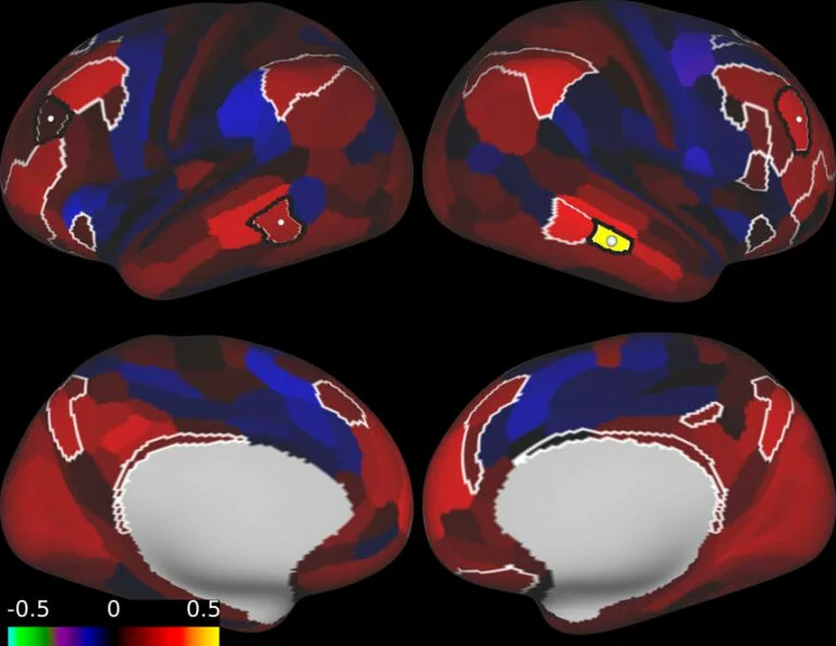
According to the National Institutes of Health, functional MRI measures changes in blood oxygenation associated with neural activity.
Resting-state functional MRI specifically evaluates how different regions of the brain communicate while the patient is not performing a task.
This allows clinicians to analyze:
At Neurotherapeutix, this data becomes the foundation for computational analysis and personalized treatment planning.
Computational brain mapping may be appropriate for individuals experiencing psychiatric, neurological, or cognitive symptoms that involve disrupted neural communication.
Some patients pursue computational brain mapping after traditional treatments have not produced adequate improvement. Others seek advanced imaging to better understand the neurological basis of persistent symptoms.
Because many conditions involve network dysfunction rather than isolated structural abnormalities, functional connectivity analysis can provide valuable insight into how the brain is functioning.
Brain mapping is commonly used in patients experiencing:
For individuals exploring TMS therapy for depression or related psychiatric care, computational brain mapping may help identify the neural circuits most closely associated with symptoms.
This information can support more individualized treatment planning and targeting.
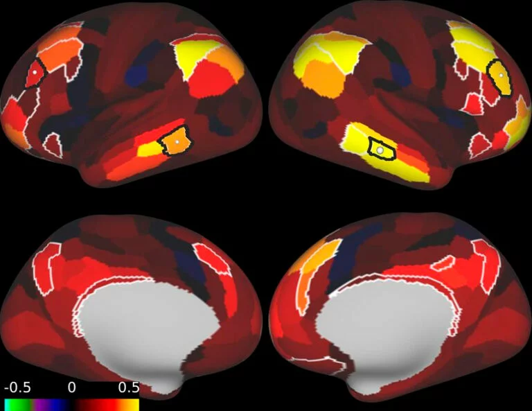

Brain mapping may also help evaluate patients experiencing:
Because symptoms may persist even when structural scans appear relatively stable, functional imaging can provide additional insight into how neural networks have been affected.
Computational brain mapping is especially valuable for patients considering fMRI-guided TMS therapy.
Traditional TMS approaches often rely on generalized anatomical positioning. At Neurotherapeutix, clinicians use connectivity analysis to personalize stimulation targets around each patient’s unique neural network activity.
This precision-guided approach helps differentiate personalized TMS vs. standard TMS protocols by focusing on functional brain activity rather than generalized landmarks alone.

At Neurotherapeutix, computational brain mapping combines advanced imaging, analysis, and individualized treatment planning.
The goal is not simply to create images of the brain, but to better understand how neural systems communicate and how those connectivity patterns may relate to symptoms.
Neurotherapeutix is the only clinical practice in the United States using patented fMRI-guided computational brain mapping to personalize TMS therapy and neurological treatment planning. This approach was developed to move beyond generalized anatomical targeting and toward individualized connectivity-based care.
During the rsfMRI scan, patients lie comfortably inside the MRI scanner while the brain’s resting connectivity patterns are recorded.
Unlike task-based imaging, resting-state fMRI does not require patients to complete cognitive exercises during the scan.
Traditional task‑based fMRI only measures brain activity during simple, predefined tasks (like moving a hand or looking at pictures), giving a narrow view of isolated circuits.
Resting‑state fMRI, by contrast, maps the brain’s intrinsic network activity. At the same time, you simply lie in the scanner, generating a whole‑brain connectome that can reveal subtle disruptions in motivation, mood, attention, and other symptom‑related networks that task‑based scans often miss.
The imaging process measures blood oxygen level-dependent (BOLD) signals across neural systems to evaluate how brain regions naturally coordinate activity.
According to the National Institute of Biomedical Imaging and Bioengineering, MRI technology does not use ionizing radiation and is considered non-invasive.
Many patients tolerate the scan well and can return to normal activities afterward.
After imaging is completed, the data undergoes advanced computational analysis.
Clinicians evaluate:
Rather than focusing solely on isolated regions, the analysis examines how networks interact across the entire brain.
This network-based approach helps clinicians better understand the functional systems associated with psychiatric and neurological symptoms.
Patients interested in imaging comparisons can also explore rsfMRI vs. SPECT imaging.
The information gathered through computational brain mapping becomes the foundation for personalized treatment planning.
Two patients with the same diagnosis may have very different connectivity patterns and symptom drivers. This is one reason individualized analysis matters.
At Neurotherapeutix, clinicians use rsfMRI data to identify the neural circuits most closely associated with each patient’s symptoms before treatment begins.
This information may help guide TMS therapy targeting, monitor network changes over time, and support more precise clinical decision-making.
fMRI-guided TMS therapy depends on accurate targeting of the neural circuits involved in symptoms.
At Neurotherapeutix, computational brain mapping helps clinicians determine where to deliver stimulation and how to personalize treatment for each patient.
Patients interested in the overall treatment process can also learn more about how TMS therapy works.
Many psychiatric and neurological symptoms involve disrupted communication across interconnected brain systems.
By evaluating connectivity patterns before treatment, clinicians can personalize stimulation targets around:
Rather than relying solely on generalized positioning, treatment is guided by the patient’s individual brain function.
Computational brain mapping may also help clinicians evaluate changes in connectivity patterns over time.
As treatment progresses, imaging and clinical response can provide insight into how neural networks are adapting and reorganizing.
This focus on functional change supports a more personalized, data-informed treatment process.
Computational brain mapping using rsfMRI is non-invasive and does not involve ionizing radiation.
Patients remain awake throughout the scan and do not require sedation in most cases.
During the appointment:
The overall appointment length may vary depending on the imaging protocol and evaluation process.
Because MRI technology uses magnetic fields rather than radiation, rsfMRI is considered a safe imaging modality when appropriate screening guidelines are followed.
Computational brain mapping helps clinicians evaluate how neural networks communicate and function together. At Neurotherapeutix, rsfMRI-based brain mapping is used to support personalized treatment planning for psychiatric and neurological conditions.
Appointment times vary depending on the imaging protocol and consultation process. Your care team will provide individualized guidance before the evaluation.
Neurotherapeutix is a private-pay practice. We do not submit insurance claims or communicate directly with insurance companies. However, patients may receive documentation, clinical records, and billing information to independently pursue reimbursement if they choose to do so.
Yes. At Neurotherapeutix, computational brain mapping is the first step in the treatment process.
Using rsfMRI and patented computational analysis, clinicians evaluate your brain’s unique connectivity patterns before developing a personalized treatment plan.
This information helps identify the neural networks associated with your symptoms and guides individualized TMS therapy targeting.
Neurotherapeutix goes beyond standard TMS by using its cutting‑edge, patented functional brain‑mapping technology before every course of treatment—and again after treatment—to objectively measure brain‑network change and guide the next best step in each patient’s care.
Traditional MRI focuses primarily on anatomy and structural abnormalities. rsfMRI brain mapping evaluates neural connectivity — how different regions of the brain communicate and coordinate activity in real time.
Understanding how neural networks communicate can provide important insight into psychiatric, neurological, and cognitive symptoms.
At Neurotherapeutix, advanced rsfMRI brain mapping combines functional imaging, patented computational analysis, and precision-guided care to better understand how each patient’s brain networks function.
As the only clinical practice in the United States offering this patented fMRI-guided computational brain mapping approach, Neurotherapeutix provides a level of personalization not available through standard TMS protocols.
Whether you are exploring TMS therapy, neurological rehabilitation, or advanced psychiatric treatment, brain mapping may help clinicians better understand the neural circuits associated with your symptoms.
To learn more or schedule an evaluation, please contact us.

Call us at (917) 388-3090 or click to request a regular or telehealth appointment.
Neurotherapeutix
171 East 74th Street, Unit 1-1 New York, NY 10021

Neurotherapeutix is the leading clinic for functional imaging guided transcranial magnetic stimulation (TMS), a safe, innovative, and non-invasive methodology for treating a wide range of acute and chronic mental disorders and brain injuries. Our advanced fMRI technology allows us to map the brain for the… Learn More »
By: Neurotherapeutix NYC
Reviewed By: Marta Moreno, Ph.D
Published: March 24, 2023
Last Reviewed: March 5, 2026
QUICK INQUIRY
Contact us to get an estimate for your medical services requirements. You can fill in the form to specify your medical requirements or you can call us directly.


© 2024. All Rights Reserved
