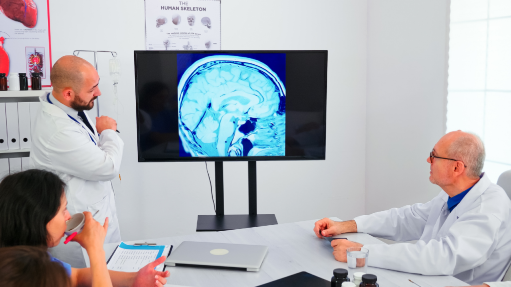
rsfMRI vs SPECT Imaging
What is resting state functional MRI or rsfMRI?
Functional MRI (fMRI) is a medical imaging technique that its signal is based on blood-oxygen level dependent (BOLD). When used to image the brain, fMRI gives us the amount of oxyhemoglobin and deoxyhemoglobin that reflects neuronal activity. Resting state fMRI (rs-fMRI) is a highly advanced method that measures the brain functional activity while the subjects are awake and lying down at rest in the scanner without performing any specific task. Because MRI has very high resolution, it is capable of producing a high-quality image of the brain and has a high contrast between gray and white matter.
rsfMRI is performed by MRI scanners that are widely available in hospitals and imaging centers. Furthermore, rs-fMRI is easy to perform and has an excellent safety record compared to other imaging modalities like SPECT and PET. Unlike SPECT and PET, there is no radiation involved in rsfMRI. rsfMRI has a resolution of the order of millimeter while SPECT has a spatial resolution of centimeter scale. This mean that rsfMRI’s resolution is 10 times better than SPECT. Furthermore, the temporal resolution of rsfMRI is of the order of 1 second while SPECT has a temporal resolution of about 15 minutes. This superior temporal resolution (time sensitivity) of rsfMRI makes it highly valuable to detect brain activity with sub-second accuracy.
Which one, rsfMRI or SPECT, is better for psychiatric disorders?
So, rsFMRI vs SPECT, which is better for TMS? As it was mentioned above, the temporal resolution associated to rsfMRI, which tells us how often brain function is measured, plays a huge role in accurately monitoring brain activity to find the malfunctioning brain regions the size of a grain of lentil. SPECT gives an image from 10-15 minutes of activity at a time, our rsfMRI can give a second-by-second picture of your brain activity. Since brain networks are made up of regions that must work in perfect harmony, sampling their activity second-by-second is vital, and rsfMRI is the method of choice.

rsfMRI for Brain Mapping Therapy
Another indispensable advantage of rsfMRI is that during the same scan, high resolution anatomical MRI images are also obtained and used to inform the functional maps. This makes rsfMRI a uniquely capable technology for identifying unique patterns of brain function, showing which parts of your brain have been affected by a psychiatric disorder. Our network approach looks at many brain regions to construct the networks and determine if brain regions such as the dorsolateral prefrontal cortex, the thalamus, putamen, basal ganglia etc., are working out of pace with their counterparts in their network. Whether a brain region is working too fast (hyperactive) or too slow (hypoactive) is an extremely valuable information that allows us to design a personalized TMS treatment to target those regions. This is a vital information about the inner working of your brain that could be used before and after treatment to assess treatment’s response. Our patients have found the pre-/post-treatment comparison at the end of their treatment period extremely gratifying and reassuring that their condition has been effectively treated and the “functional injury” to their brain is repaired.
SPECT Imaging to Confirm a Diagnosis
Conditions such as stroke, brain tumors and MS are very easy to see, and SPECT offers a faster option. Since a SPECT scan is typically shorter than fMRI, if the purpose is just to verify the diagnosis, then SPECT will do the job. But for pinpointing malfunctioning regions of your brain for accurate diagnosis of the disorder and proper treatment, rsfMRI provides a superior quantitative and clinically relevant information, which can be used for diagnosis and treatment alike.


