Contact Us

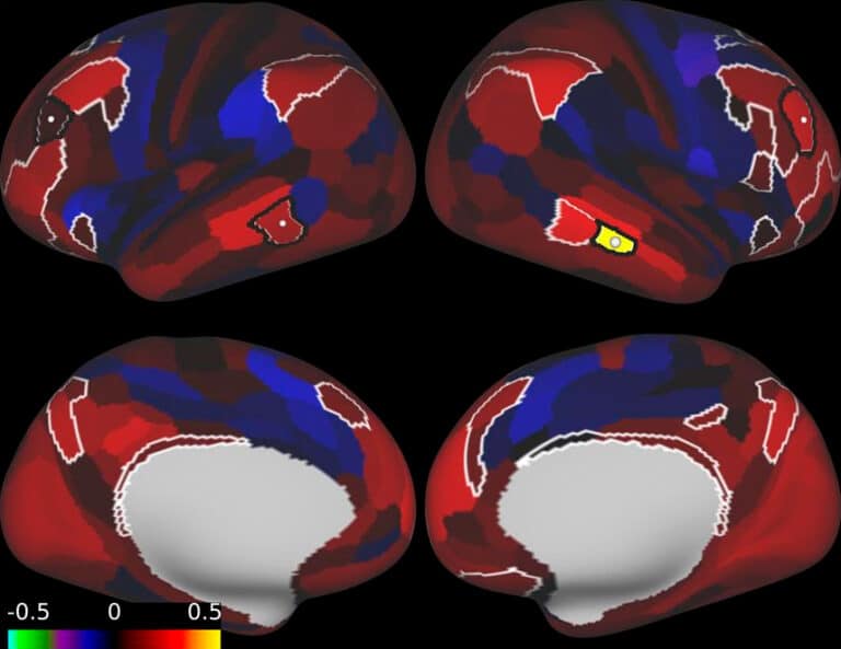
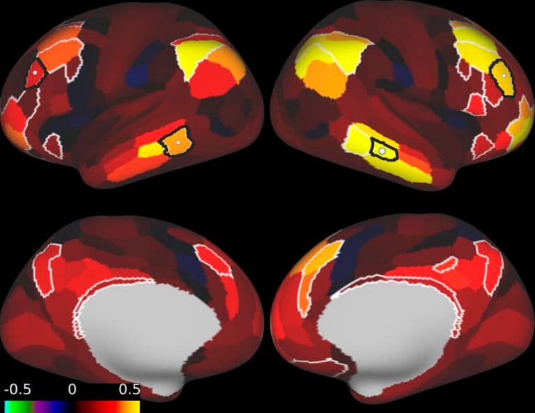
Resting-state fMRI (rsfMRI) is an imaging technique that detects functionally linked brain regions associated with various conditions. If multiple fast MRI images are acquired, then a map of fMRI can be constructed by finding the temporal fluctuations in the time series of each brain region (voxel).
Functional connectivity is then calculated by finding statistical dependencies between different regions (voxels). For Gaussian distribution of data, second-order dependencies (i.e., covariances or correlations) are found to build functional connectivity maps.
This is achieved by rsfMRI’s sensitivity to spontaneous blood oxygenation level-dependent (BOLD) contrast fluctuations. Since the BOLD signal between different brain regions that work together is temporally correlated when the subject is at rest (the subject is presented with no specific stimulus or task) rsfMRI can reveal deficiencies of various brain networks. Resting-state networks (RSN) are made of a set of brain regions with coherent spontaneous fluctuations in activity. rsfMRI allows us to explore the brain’s functional organization and to examine its differences with normal controls to find malfunctions in neurological or psychiatric diseases.
Since its invention in 19952, fMRI has revealed a number of brain networks such as default mode network (DMN), Salient Network (SN), and Central Executive Network (CEN) which are regularly found in healthy subjects and exemplify specific patterns of synchronous activity between specific brain regions.


In our work, we use behavioral, physiological and neurological evidence relevant to these networks to make our diagnosis and plan treatments. Our use of functional connectivity has been highly effective from a clinical perspective.
This is confirmed by multiple evidence regarding spontaneous brain activity gathered in the last several years, and its importance as a very sensitive biomarker of the health of the brain. DMN is one of the most important RSNs with a synchronized activity pattern indicative of its engagement with other networks.
The pattern of interaction of these major networks has been found to be associated with cognitive performance and plays an important role in neuroplasticity. Since neuroplasticity is at the core of consolidation and maintenance of brain function, any abnormality in the performance of individual networks or their interaction will constitute a distinct biomarker for a distinct disorder.
Transcranial magnetic stimulation (TMS) is a non-invasive (does not enter your body) brain stimulation technique that is used to change brain activity and correct abnormal activity due to illness, using a magnetic field. This magnetic field can pass through the skull to your brain, and induce an electric current at the site of stimulation (focal).

TMS can modulate the resting-state activity of the brain and fine-tune DMN plasticity; the direction (increase or decrease in activity) and the extent of this modulation depends on designing a specific rTMS protocol for each individual patient’s brain activity. Our work in clinical and fundamental neuroscience has been essential in improving the technology and its integration with brain-imaging methodologies such as rsfMRI.
We offer existing clinical applications of rsfMRI guided TMS therapy for many psychiatric and neurological diseases and a sample of our work on emerging possibilities such as in traumatic brain injury, dementia and stroke is available on this site.
The concept of functional synchrony on which fMRI is based was first established by Electroencephalography (EEG) which was used in the 1980’s to visualize functional connectivity in the brain1. EEG is capable of detecting long-range EEG coherence across cortical regions and between the hemispheres which makes it an important tool for monitoring functional connectivity. In this regard, EEG has powerful features such as detection of occipital alpha rhythm that offers another tool for treatment.
However, since the coherencies among nodes of brain networks are spread throughout the whole cortex and their localization is profoundly important, a technique with sensitivity over the whole brain is needed to treat patients with damage to deep brain regions.

This gives fMRI an advantage as it detects signals from the whole brain at a very high spatial resolution (mm resolution), including deep thalamic regions that are intimately involved in neuropsychiatric disorders. EEG can detect cortical (surface) regions and guide TMS therapy with its superior temporal resolution of millisecond range, which is close to the temporal activity of neuronal electrical signals, but due to its lower spatial resolution (i.e., inverse problem in EEG), is unable to identify where a particular type of activity originates in the brain (poor spatial resolution), making EEG incapable of fine-tuning specific cortical regions in need of treatment.
The temporal resolution of fMRI, on the other hand, is based on the time-scale of hemodynamic response which is about one second.
This enables fMRI to detect coherent BOLD activity in very low-frequency range, i.e., <0.1 Hz, which although not directly associated with the EEG activity in this frequency range, universal observation of BOLD activity in <0.1 Hz range has turned it into a powerful tool for the depiction of brain networks3.
Furthermore, it has been shown that there is a correspondence between BOLD and EEG, low-frequency BOLD signal fluctuations track spontaneous changes in the main EEG rhythms. At the heart of this relationship lies the role of local field potentials (LFP) in both BOLD and EEG signals that points to the common neurophysiological roots of the two techniques.
We use both BOLD (which is due to low-frequency LFPs and low-frequency modulations of high-frequency LFPs) and EEG which detects high-frequency LFPs directly to guide our TMS treatments.
The DMN is a sensitive brain feature that we use to diagnose and treat neuropsychiatric disorders. This is because DMN has an activity pattern that inversely correlates with arousal and effort.
DMN responsiveness to arousal or external stimulation also contains precious information about other networks because it interacts intimately with them. We use this powerful tool as a sensitive state-of-art therapeutic technique for psychiatry and neurology.
View sources here: https://neurotherapeutixnyc.com/sources/

Call us at (917) 388-3090 or click to request a regular or telehealth appointment.
Neurotherapeutix
171 East 74th Street, Unit 1-1 New York, NY 10021

Neurotherapeutix is the leading clinic for functional imaging guided transcranial magnetic stimulation (TMS), a safe, innovative, and non-invasive methodology for treating a wide range of acute and chronic mental disorders and brain injuries. Our advanced fMRI technology allows us to map the brain for the… Learn More »
By: Neurotherapeutix NYC
Reviewed By: Marta Moreno, Ph.D
Published: March 24, 2023
Last Reviewed: September 27, 2024
QUICK INQUIRY
Contact us to get an estimate for your medical services requirements. You can fill in the form to specify your medical requirements or you can call us directly.