


If you’re exploring brain imaging or advanced treatment options, you may come across the terms fMRI vs MRI. While they sound similar, they provide very different types of information about the brain.
An MRI shows what the brain looks like. An fMRI shows how the brain works.
You may also hear the term rsfMRI (resting-state fMRI). This is not a separate type of scan, but a specialized form of fMRI that measures how brain networks communicate at rest.
Understanding the difference between MRI, fMRI, and rsfMRI can help you make more informed decisions about your care, especially if you are considering brain-based treatments like TMS therapy.
Each imaging method plays a different role, and knowing how they work together can provide a clearer picture of your brain health.
Contact us directly or continue reading to learn more.
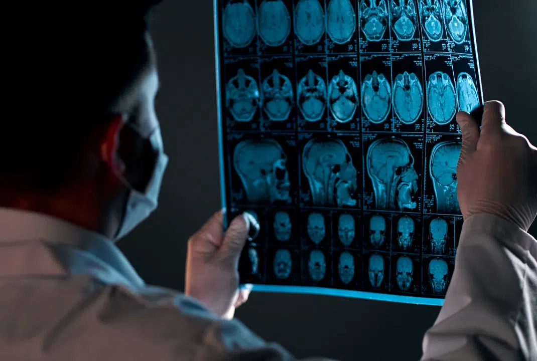
What is an MRI?
Magnetic resonance imaging (MRI) is one of the most widely used imaging tools in modern medicine.
According to the National Institute of Biomedical Imaging and Bioengineering, MRI is a non-invasive imaging technology that produces detailed anatomical images using magnetic fields and radio waves, without exposing patients to ionizing radiation.
In brain imaging, MRI plays a critical role in helping clinicians understand how the brain physically appears.
It provides highly detailed images of brain structure, making it essential for diagnosing and monitoring a wide range of neurological conditions.
How MRI works
MRI uses powerful magnetic fields and radio waves to generate highly detailed images of the body’s internal structures.
Unlike CT scans or X-rays, MRI does not use ionizing radiation, making it a safe, non-invasive option for many patients.
During the scan, the MRI machine detects signals from hydrogen atoms in the body. These signals are processed to create precise, layered images of tissues, allowing clinicians to examine structures in great detail.
What MRI shows: Brain structure and anatomy
MRI is especially effective at visualizing brain structure. It can show:
- The size and shape of different brain regions
- The integrity of brain tissue
- Structural abnormalities such as tumors, lesions, or bleeding
Because MRI focuses on anatomy, it provides critical information about physical changes in the brain. However, it does not reveal how different parts of the brain function or interact with one another.
Common clinical uses of MRI
MRI is used across many areas of medicine due to its ability to capture detailed structural images.
In brain health, it is commonly used to:
- Diagnose neurological conditions
- Evaluate traumatic brain injuries
- Detect tumors or vascular abnormalities
- Monitor disease progression over time
While MRI is a powerful diagnostic tool, it has limitations in understanding conditions that involve brain function rather than structure.
What is an fMRI?
Functional magnetic resonance imaging (fMRI) builds on the same technology as MRI but focuses on a different question: not what the brain looks like, but how it works.
This distinction is important because many psychiatric, neurodevelopmental, and neurological conditions are not caused by visible structural damage. Instead, they are related to how brain regions communicate and function together.
How fMRI measures brain activity
fMRI measures brain activity by detecting changes in blood oxygen levels, known as blood-oxygen-level-dependent (BOLD) signals.
When a specific area of the brain becomes active, it requires more oxygen. This change in oxygen levels alters the magnetic properties of the blood, which fMRI can detect and map in real time.
By tracking these changes, fMRI provides insight into which brain areas are active and how they respond under different conditions.
What fMRI shows: Function, connectivity, neural networks
Unlike MRI, fMRI reveals how the brain functions as a system.
It can show:
- Which regions are active during certain tasks
- How different areas of the brain communicate
- How neural networks support mood, cognition, and behavior
This makes fMRI particularly useful for understanding conditions involving disrupted connectivity, where the issue lies not in structure but in how the brain’s networks interact.
What is rsfMRI?
Resting-state functional MRI (rsfMRI) is a specialized form of fMRI that measures brain activity while you are at rest, without performing a specific task.
Instead of focusing on isolated regions, rsfMRI analyzes how entire brain networks communicate with one another. This provides a more comprehensive view of functional connectivity.
At Neurotherapeutix, rsfMRI is used as part of fMRI-guided computational brain mapping. This allows clinicians to identify patterns of brain activity that may be contributing to symptoms and to design more personalized treatment approaches.

Each imaging method provides a different layer of information. MRI offers structural clarity, while fMRI and rsfMRI provide insight into how the brain functions and communicates.
At Neurotherapeutix, we combine cutting-edge resting-state fMRI (rsfMRI) with structural MRI to identify abnormal brain activity down to the millimeter. This dual approach gives us both precise anatomical detail and deep functional insight, ensuring the highest level of accuracy for our computational brain mapping.
Why fMRI matters for brain-based treatment
For many patients, especially those with psychiatric disorders, understanding brain function is essential for effective treatment. This is where fMRI plays a critical role.
Limitations of structural MRI for psychiatric conditions
In many psychiatric and neurological conditions, the brain may appear structurally normal on an MRI scan. This can make it difficult to identify the root cause of symptoms solely with structural imaging.
Conditions such as depression, anxiety, post-traumatic stress disorder (PTSD), and even mild concussions are often associated with disruptions in neural connectivity, not visible structural damage.
As a result, MRI may not provide enough information to guide treatment decisions in these cases.
Furthermore, functional brain imaging allows us to detect the early stages of neurodegenerative diseases before any physical symptoms or structural damage appear. Identifying these issues early enables us to intervene sooner, maximizing the effectiveness of the treatment.
How fMRI guides precision TMS therapy at Neurotherapeutix
TMS therapy relies on accurate targeting of the brain circuits involved in symptoms. The more precisely these circuits can be identified, the more personalized the treatment can be.
At Neurotherapeutix, we use fMRI-guided TMS therapy to analyze brain activity and guide stimulation. By using rsfMRI data, we can:
- Identify specific neural networks involved in symptoms
- Tailor treatment to individual brain connectivity patterns
- Adjust treatment as brain function evolves over time
This precision-driven approach allows for more individualized care compared to traditional methods that rely on standardized targeting.
fMRI-based computational brain mapping
Using computational brain mapping, clinicians can visualize how brain networks interact and change over time.
This approach supports:
- More informed treatment planning
- Ongoing evaluation of progress
- A deeper understanding of brain function
By combining advanced imaging with personalized care, this method helps bridge the gap between diagnosis and treatment.
Learn more about advanced brain imaging at Neurotherapeutix
Understanding the difference between fMRI and MRI is an important step in exploring advanced, brain-based care options.
If you’re considering treatment or want to better understand how brain imaging can support your care, you can explore our comparison of rsfMRI vs SPECT imaging to see how different technologies are used in clinical practice.
At Neurotherapeutix, we use functional imaging to guide personalized, non-invasive medical services for a full range of mental health and neurological conditions.
If you’re ready to learn more, you can request an appointment to speak with our team.
FAQs: fMRI vs MRI
What is the main difference between MRI and fMRI?
MRI shows the structure of the brain, while fMRI shows how the brain functions. MRI focuses on anatomy, whereas fMRI measures activity and connectivity.
What is rsfMRI, and how is it different from a regular fMRI?
rsfMRI is a subtype of fMRI that measures intrinsic brain activity at rest. While traditional task-based fMRI is limited by the specific task used and requires the patient’s active collaboration, rsfMRI overcomes these limitations. It provides a task-independent, whole-brain analysis of functional network connectivity, making it an ideal, unbiased method for personalized treatment planning.
What does an fMRI scan measure?
An fMRI scan measures changes in blood oxygen levels associated with neural activity. These changes provide insight into which brain areas are active and how different regions interact.
Is fMRI safe? How long does it take?
Yes, fMRI is non-invasive and does not involve radiation. Most scans take between 30 minutes and one hour, depending on the type of imaging being performed.
Why is fMRI important for TMS therapy?
TMS therapy depends on accurate targeting of brain circuits. fMRI provides detailed information about brain activity and connectivity, allowing clinicians to personalize treatment rather than relying on generalized targeting.
At Neurotherapeutix, this approach is taken a step further: it is the only clinical practice in the United States using fMRI-guided computational brain mapping to guide TMS therapy, enabling a more precise and individualized treatment strategy.


